Gastrointestinal Radiology
1. Recognize the different parts of the gastrointestinal tract.
2. Trace the blood flow from the aorta to the digestive organs in angiograms.
3. Become familiar with ultrasound using images of the gallbladder and kidney.
4. Trace the flow of blood in the hepatic portal system using a portogram.
In a frontal (AP) view of the abdomen, note the outline of the lateral edge of the psoas muscle. The kidney and psoas muscles can be seen because retroperitoneal fat, which allows a greater passage of x-rays than the tissue of the kidneys and muscle, surrounds these structures. The right kidney is usually seen moderately lower than the left because of the overlying liver. Swallowed air can be seen in the stomach in this film. The position of the inguinal ligament, extending between the anterior superior iliac spine and the pubic tubercle, has been drawn on the left. Phleboliths (calcified blood clots) in pelvic veins are seen; this is a variation from normal in older patients and not pathologic.
An axial CT scan at the level of the body of L4 is shown. The small intestine has been opacified with swallowed barium contrast material which has not yet reached the colon. Sections of the colon can be seen filled with air and fecal material. Intravenous contrast material has outlined the proximal common iliac arteries as well as the inferior vena cava to the patient's right of these arteries. Note the prominent fat about the psoas muscles. As indicated above, fat, with a different x-ray density, appears dark as it surrounds the more radiopaque psoas muscles. The muscles of the abdominal wall are also seen.
Swallowed barium contrast outlines the position of the stomach, duodenum, jejunum, and ileum in relation to abdominal quadrants. Note the stomach is largely in the left upper quadrant, the duodenum is in the right upper quadrant, the jejunum is in the left upper and left lower quadrants, and the ileum is mostly in the right lower quadrant. The position of the umbilicus is shown in relation to the transcristal line (line across the iliac crests) and the midline. This intersection is usually at the level of L4. A barium enema (below) with reflux of barium into the ileum is shown, and the quadrants are again indicated. The cecum is in the right lower quadrant; hepatic flexure in the right upper quadrant; splenic flexure in the left upper quadrant; and the descending colon is in the left upper and left lower quadrants. The sigmoid colon usually lies in the left lower quadrant and the rectum in the midline. The appendix is not seen in this study, but it is almost always in the right lower quadrant.

MRI and magnetic resonance angiography (MRA) can be used to study the major abdominal arteries. In the sagittal plane shown, note the level of origin of the celiac trunk and superior mesenteric artery (SMA). The MRA shows the left renal artery in cross-section immediately posterior to the SMA and anterior to the abdominal aorta. These vary between T12 and L1. Under fluoroscopic guidance, catheters can be placed in the celiac trunk or the superior mesenteric artery for injection of contrast materials. The images here were made following the injection of contrast material (gadolinium) in an antecubital vein. The contrast material flowed into the systemic circulation through the heart and lungs.
Study the selective angiogram of the celiac trunk. Note the tortuous course of the splenic artery as it goes toward the spleen. The right and left gastric and gastroepiploic vessels can be seen. The latter vessels course along the greater curvature of the stomach; this patient's stomach has an unusually low position. Note the characteristic downward course of the gastroduodenal artery as it passes behind the first part of the duodenum. The pancreaticoduodenal arteries are not well seen.
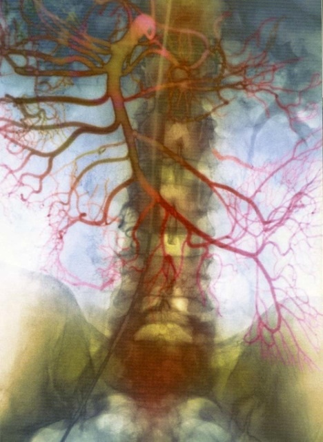
Examine the angiogram of the superior mesenteric artery, giving attention to the point of origin from the aorta at L1. The inferior pancreaticoduodenal branch can also be seen but it is faint. The middle, right, and ileocolic arteries are well defined. Note the formation of arterial loops in the mesentery. The branches supplying the small bowel emerge from the loops as vasa rectae. Contrast material can be seen in the right ureter because of renal excretion from a preceding celiac artery injection.
Two prone PA projections of the stomach and duodenum are shown. In figure 1, the stomach is almost completely distended with a barium meal. Note the divisions of the stomach, including the cardia, fundus, body, and pyloric antrum. The cardia is adjacent to the esophageal junction with the stomach, and the fundus is the area superior to the cardia that fills with swallowed air in the upright position. Peristaltic waves can be seen on both the lesser and greater curvatures of the stomach. They can appear quite prominent when observed fluoroscopically. The pyloric canal is very short and is surrounded by the pyloric musculature. In figure 2, peristalsis has largely emptied out the lower aspect of the stomach and linear streaks of barium can be seen between the gastric rugae. In this figure the duodenum is well outlined. The first part of the duodenum (bulb) is usually at the level of L1, the second (descending portion) is at L2, the third portion crosses L3, and the fourth ascends to the ligament of Treitz on the left side of L2. In this patient the duodenum is shifted slightly to the right, however. The head and medial part of the body of the pancreas lie within the C-shaped loop. Mucosal folds in the duodenum and the jejunum vary from striations to irregular dark shadows in the barium.
A transverse MRI section including the pancreas and adjacent organs is compared with a CT transverse section at a slightly lower level. In the MRI, the lobular configuration of the pancreas can be seen. Note the relationship of the head of the pancreas within a cross-section of the duodenal loop. The splenic vein in this scan is immediately posterior to the pancreas as it drains the portal vein. The superior mesenteric artery with contrast material can be seen in the CT section between the pancreas and the aorta. The left renal vein can be observed crossing the aorta posterior to the superior mesenteric artery. Prior to the development of CT and MRI scanning, the pancreas could not be imaged well in vivo. Now pancreatic cancer and also pancreatitis can be diagnosed fairly accurately with the aid of these techniques.
Using a CT or MR image at this level, one should be able to appreciate:
a) The head of the pancreas lying within the C-shaped part of the duodenum. The duodenum is to the cadaver's right of the head.
b) The tail of the pancreas projecting toward the spleen.
c) The ventral surface of the pancreas indicating the posterior wall of the lesser omental bursa. Note that the anterior wall of this bursa is the posterior wall of the stomach.
e) The superior surface closely associated with the splenic vessels.
d) The pancreatic incisure occupied by the superior mesenteric vessels. The left renal vein can easily be seen coursing across the aorta.
Another way to outline the portal system is by direct injection of contrast material via a catheter placed in the portal vein extending to the splenic vein. In the study shown, the catheter was initially installed in the liver by inserting a needle between the ribs. The catheter was passed through the needle into the portal vein and eventually the splenic vein by observing its passage with a fluoroscope. The superior mesenteric vein is not seen because of non-opaque venous blood flowing out of it. This study was done prior to the placement of a conduit between the portal vein and inferior vena cava.
Examine a frontal abdomen film made 15 minutes after injection of intravenous contrast material. The iodinated contrast material has been filtered through the kidneys and opacifies the calyces, pelves, ureters, and bladder. The right kidney is slightly lower than the left due to the overlying liver. Note the proximity of the 11th and 12th ribs to the superior aspect of the kidneys. There are three natural narrowings of the ureters: (1) where the ureters begin at the renal pelves, (2) where they cross the iliac vessels, and (3) at the entry point of the ureters into the bladder (ureto-vesicle junction). The slightly sinuous outline of the ureters is due to peristaltic activity. The indentation of the superior wall of the bladder is due to the uterus in this patient. Observe the medial border of each kidney lying along the lateral margin of the "psoas shadow".
Study the coronal and axial MRI sections of the kidneys following the injection of intravenous contrast material. The renal cortices as well as columns are opacified. The renal pyramids stand out between these structures. In the axial section, the left renal vein is seen between the aorta and proximal superior mesenteric artery. Note the prominent retroperitoneal fat around the kidneys in the coronal section. The left kidney appears smaller than the right because the kidneys were not in the same antero-posterior position in this plane.