Musculoskeletal Radiology
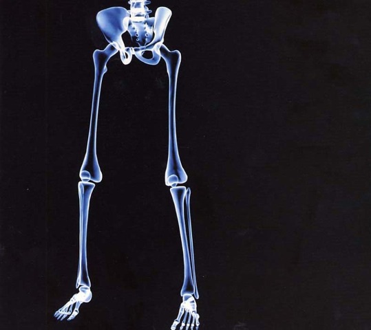
The Lower Limb
1. Identify the major bones of the hip, legs, knee, ankle and foot.
2. Locate and identify the major arteries in the hip, legs, knee, ankle and foot.
3. Interpret and translate a radiograph of the hip, knee foot and ankle.
A femoral arteriogram in a child is shown. Contrast material was injected in the lower abdominal aorta through a catheter introduced in the left femoral artery. The femoral artery with its division into a deep branch and its continuing branch is shown. Note the course of the medial femoral circumflex artery as it curves around the insertion of the iliopsoas muscle. The medial circumflex artery supplies the majority of blood to the femoral head and neck, while the lateral femoral circumflex provides some blood to the femoral head via an ascending branch and blood to muscles on the lateral side of the thigh via a descending branch.
Examine an AP film of the hip joint. Note the space between the bony outlines of the femoral head and the acetabulum. This space is occupied by the articular cartilage of the femoral head and acetabulum as well as the acetabular labrum. A focal curvilinear space (fovea capitis) in the medial aspect of the femoral head can be seen. This is the site of attachment of the round ligament (ligamentum teres). The intertrochanteric area is a common site for hip fractures. Also note the curving line formed by the superior border of the obturator foramen and the underside of the neck of the femur. This is called "Shenton's Line," and if the femur were dislocated, continuity of this line would be broken.

Examine the two axial CT sections of the hips and acetabula. The acetabulum is best depicted in axial CT sections. CT is especially helpful in hip fractures, where a possible acetabular fracture cannot be seen in conventional radiographs. In Figure 1, the right fovea capitis can be seen, as well as the round ligament. Figure 2 shows the normal orientation of the femoral neck with the femoral head when the patient is supine. The femoral neck is directed posteriorly in an oblique direction.

AP and lateral radiographs of the knee are shown. The knee joint space is clearly seen between the femur and the tibia in these images. The articular cartilages of the femur and tibia primarily occupy this area; the medial and lateral menisci fill only a small amount of this space. In the AP projection, note the medial femoral condyle, which is slightly larger than the lateral, and the tibial spines which anchor the cruciate ligaments to the tibia. The two tibial condyles and the central aspect of the tibia between them make up the "tibial plateau". This is a common site for major fractures of the knee joint. A "sunrise view" of the patella, below, (a tangential radiograph of the knee) shows the normal relationship between this bone and the adjacent femoral condyles. The articular space between the patella and the medial femoral condyle has a steeper inclination than the contralateral side. This has an implication for subluxation of the patella, allowing for easier lateral displacement.
Two sagittal MRI images depict the anterior and posterior cruciate ligaments. Note that the anterior cruciate ligament runs in a superior and posterior direction from the tibia. The posterior cruciate ligament, on the other hand, runs in a superior and anterior direction from the tibia (at its most posterior aspect). The quadriceps femoris tendon as well as the patellar ligament are clearly seen as dark shadows superior and inferior to the patella. Tendons in general have very little signal with MRI and make dark shadows, in contrast to the gray shadow representing the cartilage around the intercondylar groove. The cartilage at the posterior aspect of the patella is also gray (same MR signal as produced by the articular cartilage of the femur).
A 4 mm thick coronal T2 MRI image of the knee brings out sections of the menisci and cruciate ligaments. In this image, the menisci have a triangular appearance. Note that the anterior cruciate ligament arises from the anterior tibial spine and the surface of the tibia immediately adjacent to it. The posterior cruciate ligament is seen in cross-section in the intercondylar groove. A sagittal MRI in another patient through the lateral aspect of the lateral femoral condyle shows a tear in the posterior aspect of the lateral meniscus.
Examine the AP and lateral arteriograms of the popliteal artery and its branches. These films of knee vessels were made within one second of contrast material injection into an ipsilateral femoral artery. The vessels seen are normal. Note the mild anterior curvature of the popliteal artery in the lateral projection. This curvature of the vessel begins as the knee is flexed. The popliteal artery begins where the femoral artery exits the adductor canal. Note the extensive collateral blood supply about the knee joint through the genicular arteries. In the AP projection, the three major arteries supplying the leg and foot (anterior tibial, posterior tibial, and peroneal) are visualized. These are frequently obstructed by atherosclerosis, which may lead to decreased oxygenation of muscles resulting in intermittent claudication (pain) as the patient walks.
A lateral view of the ankle illustrates the tibiotalar joint. The talar trochlear (dome) is seen. Note the relationship between the talus and calcaneus at the subtalar joint. The cortex of the sustentaculum tali of the calcaneus is outlined, as are the articulations between the navicular, talar, cuboid, and cuneiform bones.
A frontal view of the ankle shows the medial and lateral malleoli. These two structures, as well as the superior aspect of the talus, make up the ankle mortise.
Two coronal oblique CT sections of the ankle are shown. The more posterior section defines the bony relationships around the tibiotalar joint. The subtalar joint is also well shown. The more anterior section brings out the sustentaculum tali of the calcaneus, as well as the head of the talus. Note that both structures are on the medial aspect of these bones. CT examination of the tarsal bones is sometimes required to bring out subtle fractures as well as major displacements of fracture fragments.
A dorsoplantar (AP) film (with some obliquity) brings out the metatarsal bones and phalanges. Note the two sesamoid bones at the first metatarsophalangeal joint. These are imbedded in the tendons of the two heads of the flexor hallucis brevis.
Examine the AP and lateral arteriograms of the foot. Contrast material was introduced into the femoral artery for this study. Note the connection of the dorsalis pedis and posterior tibial arteries through their branches to form the deep plantar arch. The arcuate artery also forms an arch on the dorsal surface of the foot and gives rise to dorsal metatarsal arteries. The plantar metatarsal arteries arise from the deep plantar arch. Arteriography of the foot may be carried out to assess the status of these vessels in patients who have diabetic vasculopathy.